https://www.traditionrolex.com/46
by Jan D. Bastmeijer ~ 2016
Suwidji Wongso & Co.
 
These pages are on the cultivation and taxonomy of plants of the genus Cryptocoryne & Lagenandra (Araceae)
 
CSS MenuMaker
 
HOME > Botanical > SEM

SEM studies in Cryptocoryne and Lagenandra

Habitats Spathe Chromosomes Fine Arts Documents Synonyms Whos Who
In 1998, Marian Ørgaard & Niels Jacobsen published a study on the surface structures of the spathe in Cryptocoryne and Lagenandra. This page is a short synopsis of the article adapted for a broad audience interested in Cryptocoryne.
In Scanning Electron Microscopy (SEM), imaging is done by means of an electron beam with magnetic lenses. The specimen is, to be 'visible' for electrons, covered with an extremely thin layer of gold. You see B/W pictures, for color has no meaning in SEM. Though the magnification is not very great in the pictures below, SEM pictures have an amazing great depth of field making them very attractive.


Click on the picture to get the full image (ca 50 k)
Cryptocoryne ferruginea
A. The warty surface of the limb just above the collar. The cracks are due to the freezing in liquid nitrogen for preparation.
(idem)
Note the typical conical shape of the kettle of C. ferruginea
B. The flap (valve) over the male flowers as prolongation of the spathe tube margin
       
Cryptocoryne walkeri
A. The male flowers with the two pollen sacks. Note the alveoli ('windows') in the kettle wall opposite the flowers. B. A male flower showing the opening of the pollen sacks. The pollen is released in a rather viscous fluid. C. The female flowers at the bottom of the kettle. Note the papillous surface of the stigma's and, near the spadix, the olfactory bodies.
       
Cryptocoryne pontederiifolia
A. Top of the kettle showing the transition between the mucilage covered lower part and the trichome cells above. Note. The vertical ridges are normal in C. pontederiifolia. B. The transition area in greater detail. At top the trichome cells are seen. C. The downward pointed trichome cells in the upper part of the kettle
       
Lagenandra lancifolia
photo Bastmeijer
A. View from above on the septum (the transverse wall above the kettle). Most Lagenandra's have this structure but not all. B. Male flower with pollen released. Note the ellipsoid form of the pollen corns in the slimy fluid. C. A view on the lower part of the kettle. In front a stigma with a papillous surface and near the spadix the irregular formed olfactory bodies.
       
Cryptocoryne spiralis
Left and above. Cells in the upper part of the kettle with downward pointing trichomes, some sunken into the cell cavity. Cryptocoryne albida Left. Inner surface of the tube with partly sunken trichomes.
Above. Cells in the tube with sunken trichomes forming a lattice.
       

pictures: Marian Ørgaard and Niels Jacobsen
drawings: Line Jacobsen

Three main points in the article:
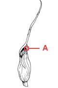
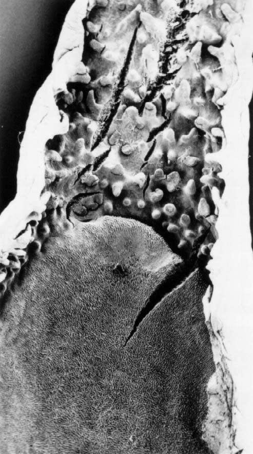
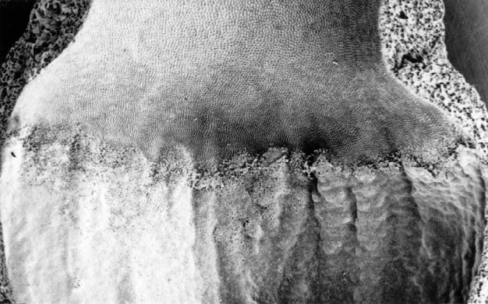
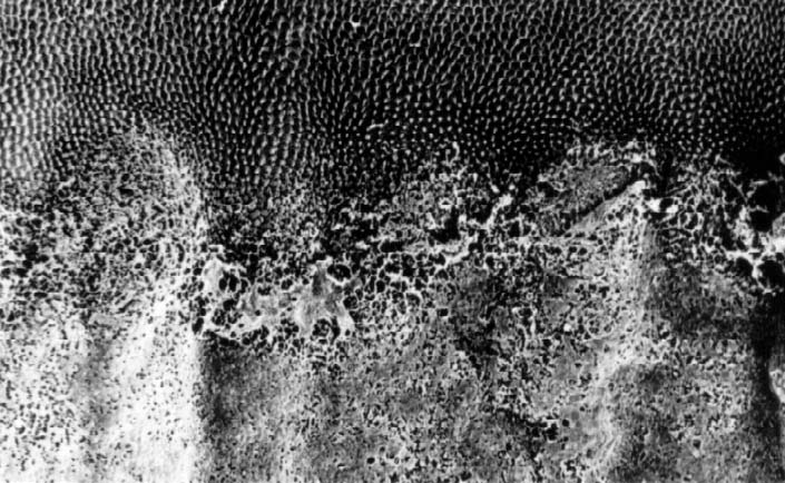
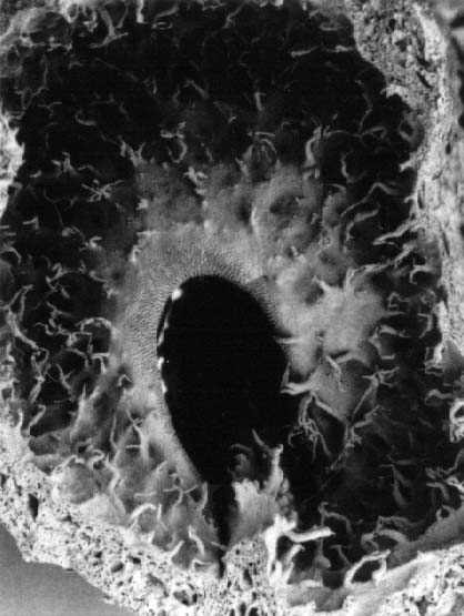
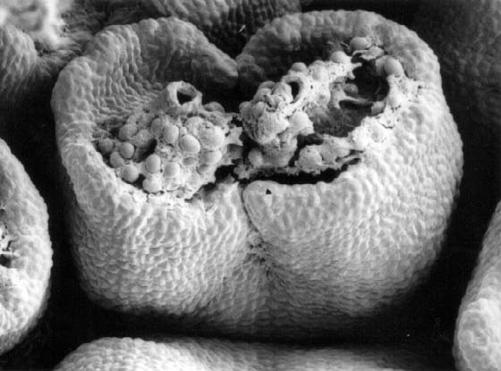
- The lower part of the kettle has a mucilage covering, interpreted as a hitherto unnoticed food source for visiting insects.
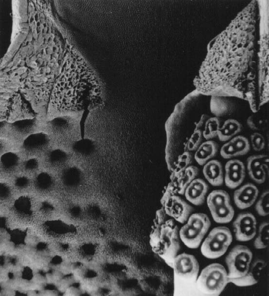
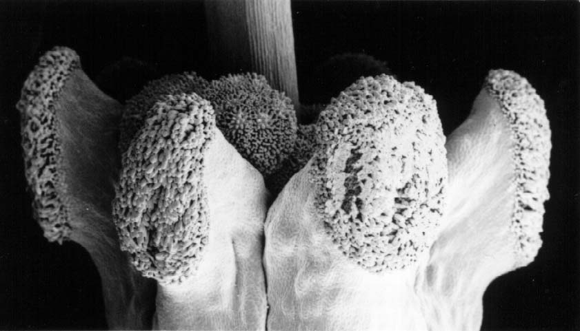
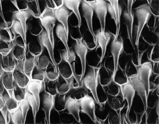
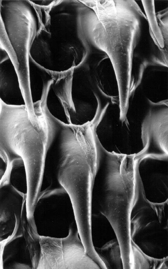
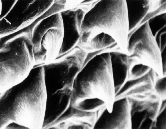
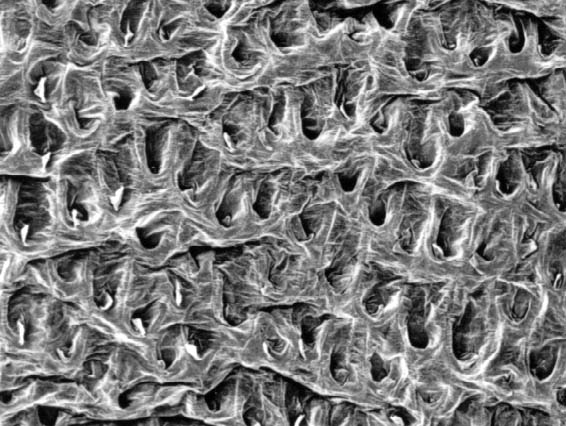
- The cells of the inner surface of the tube and the kettle have downward pointing trichomes, which collapse after two days and sink into the cell lumen. A lattice like structure remains enabling insects to climb out of the kettle and tube.
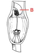
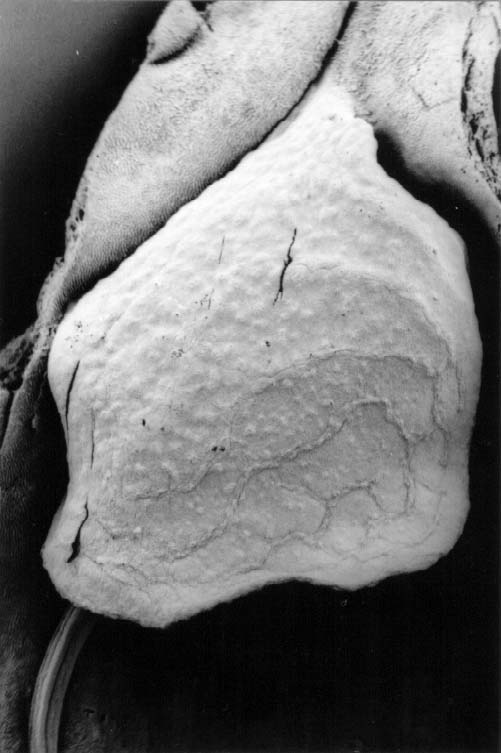
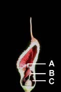
- The flap covering the male flowers is interpreted as a prolongation and continuation of the spathe tube margin.

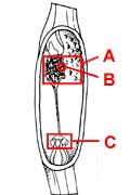
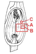
See also the page Spathe, Inside the kettle, and Foliage, rhizome and roots

The article with much more detail and discussion and 73 pictures:
Ørgaard, M. & Jacobsen, N., 1998. SEM study of surfaces of the spathe in Cryptocoryne and Lagenandra (Araceae: Aroideae: Cryptocoryneae). Botanical Journal of the Linnean Society 126: 261-289.

 
Home | Introduction | by Alphabet by Country Botanical How to | Documents | Lagenandra | Site Map 
Copyright © 2024 the Crypts pages by Jan D. Bastmeijer All rights reserved.
https://www.traditionrolex.com/46